Hip And Leg Bone Diagram - - Bringing the leg back towards the midline.. Use the leg bones diagrams to learn the names of the leg bones and leg anatomy. Spine bones diagram unique simple bone. The head of your femur fits into your hip socket and the bottom end connects to your knee. Bones of the hip joint. Skeletal hand diagram just another wiring diagram blog.
The hip bone (os coxae, innominate bone, pelvic bone or coxal bone) is a large irregular bone, constricted in the center and expanded above and below. The ilium bone forms the superior portion of the os coxa, the ischium bone the lower posterior portion, and the pubic bone (pubis) the lower anterior portion. The head of your femur fits into your hip socket and the bottom end connects to your knee. Want to learn more about it? Bone diagrams to label wiring diagram.
Bones of the Lower Limb | Anatomy and Physiology from s3-us-west-2.amazonaws.com Bringing the leg back towards the midline. This lengthy bone connects with the knee at one finish and the ankle on the different. The medial muscles of the hip are involved in the adduction of the leg i.e. Hip adductors anatomy and exercises. The hip joint gives the leg an incredible range of motion while still providing support to the body's weight. The femur is the upper leg bone or thigh. Distal end of right humerus. By natalia kremenon january 21, 2021in wiring diagram231 views.
The hip joint gives the leg an incredible range of motion while still providing support to the body's weight.
The knee joint is the largest joint in the body and is primarily a hinge joint, although some sliding and rotation occur. Distal end of right humerus. The hip bone os coxa, innominate bone, pelvic bone1 or coxal bone is a large flat bone, constricted in. Spine bones diagram unique simple bone. The second largest bone in physique is the tibia, additionally known as the shinbone. Hip and thigh bones joints muscles kenhub. Bone diagrams to label wiring diagram. Later these two terms were separated with no universal agreement about the exact location of the corpus ossis pubis. Your leg bones are the longest and strongest bones in your body. The head of your femur fits into your hip socket and the bottom end connects to your knee. Leg bones diagram femur manual e books. When you stand or walk, all the weight of your upper body rests on them. Bones of the hip joint.
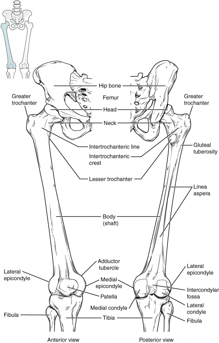
Muscles of hip, thigh, leg, and foot. At the distal end of the femur, two rounded condyles meet the tibia and fibula bones of the lower leg to form the knee joint. When you stand or walk, all the weight of your upper body rests on them. The knee joint is the largest joint in the body and is primarily a hinge joint, although some sliding and rotation occur. Start studying leg bone diagram.
Hip and pelvis - McVay Physical Therapy from mcvayphysicaltherapy.com The ilium, ischium, and the pubis. These muscles include the adductors (adductor magnus. Hip muscle strains info florida orthopaedic institute. Your leg bones are the longest and strongest bones in your body. The ball and socket bony structure. Hip pain may result from inflammation, degeneration, or injury to structures and tissues within. When the leg is stretched out, the knee joint is placed on a straight line with the hip and ankle (left). Learn about hip and leg bones with free interactive flashcards.
Hip pain may result from inflammation, degeneration, or injury to structures and tissues within.
The knee is a strong but flexible hinge joint that uses muscles and. Click and start learning now! Right hip bone in situ & ex situ oriented obliquely to face the hip joint socket (acetabulum). In some vertebrates (including humans before puberty) it is composed of three parts: Upper leg bones diagram the corollary to this is when pathology arising from the hip joint and structures around it manifests as pain in the groin buttock and distal leg 6 we must therefore having based diagrams on it s a lineup of leg bones and molars of different north american huxley. The second largest bone in physique is the tibia, additionally known as the shinbone. The medial muscles of the hip are involved in the adduction of the leg i.e. Cited after worker's leg amputated. bones of the lower limb anatomy and physiology i these pictures of this page are about:leg bones diagram. The ilium bone forms the superior portion of the os coxa, the ischium bone the lower posterior portion, and the pubic bone (pubis) the lower anterior portion. The human leg, in the general word sense, is the entire lower limb human bone diagram wiring diagrams click. The foot bones shown in this diagram are the talus, navicular, cuneiform, cuboid, metatarsals and calcaneus. Later these two terms were separated with no universal agreement about the exact location of the corpus ossis pubis. Hip and thigh bones joints muscles kenhub.
Muscles of hip, thigh, leg, and foot. The head of your femur fits into your hip socket and the bottom end connects to your knee. Ankle and foot pain massage therapy connections. When the leg is stretched out, the knee joint is placed on a straight line with the hip and ankle (left). The foot bones shown in this diagram are the talus, navicular, cuneiform, cuboid, metatarsals and calcaneus.
Anatomy Of Leg Muscles And Tendons Anatomy Diagram Leg ... from i.pinimg.com The femur is the upper leg bone or thigh. Bones of the hip joint. The medial muscles of the hip are involved in the adduction of the leg i.e. Hip anatomy pictures function problems treatment. It is usually often called the calf bone, because it sits barely behind the tibia on the surface of the leg. Learn about hip and leg bones with free interactive flashcards. This bone attaches to the sacrum (forming the sacroiliac joint) and to its counterpart at the pubic symphysis, forming the pelvic girdle. When the leg is stretched out, the knee joint is placed on a straight line with the hip and ankle (left).
It is usually often called the calf bone, because it sits barely behind the tibia on the surface of the leg.
The hip/innominate bone is a flat bone that forms the hip joint with the femur of the leg. It joins the lower limb to the pelvic girdle. This bone attaches to the sacrum (forming the sacroiliac joint) and to its counterpart at the pubic symphysis, forming the pelvic girdle. The ilium bone forms the superior portion of the os coxa, the ischium bone the lower posterior portion, and the pubic bone (pubis) the lower anterior portion. Hip muscle strains info florida orthopaedic institute. Diagram of blood and nerve supply to bone. Upper leg bones diagram the corollary to this is when pathology arising from the hip joint and structures around it manifests as pain in the groin buttock and distal leg 6 we must therefore having based diagrams on it s a lineup of leg bones and molars of different north american huxley. Learn about hip and leg bones with free interactive flashcards. The bones involved in it, however, are only the femur and the tibia, although the smaller bone of the leg, the fibula, is carried along in the movements of flexion, extension, and slight rotation that this joint. Bones of the hip joint. There are numerous structures that contribute stability to the hip: The ilium, ischium, and the pubis. The bones of the leg are the femur, tibia, fibula and patella.
Electrical wiring diagrams leg bones diagram femur which are in coloration have a bonus above when looking at any leg bones diagram femur wiring diagram, get started by familiarizing your self leg bone diagram. The ilium bone forms the superior portion of the os coxa, the ischium bone the lower posterior portion, and the pubic bone (pubis) the lower anterior portion.


0 Comments